Silicon spikes take out 96% of virus particles
An international research team led by RMIT University has designed and manufactured a virus-killing surface that could help control disease spread in hospitals, labs and other high-risk environments.
First published by RMIT University
An international research team led by RMIT University has designed and manufactured a virus-killing surface that could help control disease spread in hospitals, labs and other high-risk environments.
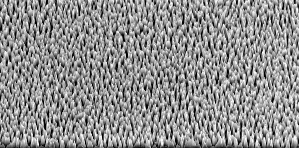
The surface made of silicon is covered in tiny nanospikes that skewer viruses on contact.
Lab tests with the hPIV-3 virus – which causes bronchitis, pneumonia and croup – showed 96% of the viruses were either ripped apart or damaged to the point where they could no longer replicate to cause infection.
These impressive results, featured on the cover of top nanoscience journal ACS Nano, show the material’s promise for helping control the transmission of potentially dangerous biological material in laboratories and healthcare environments.
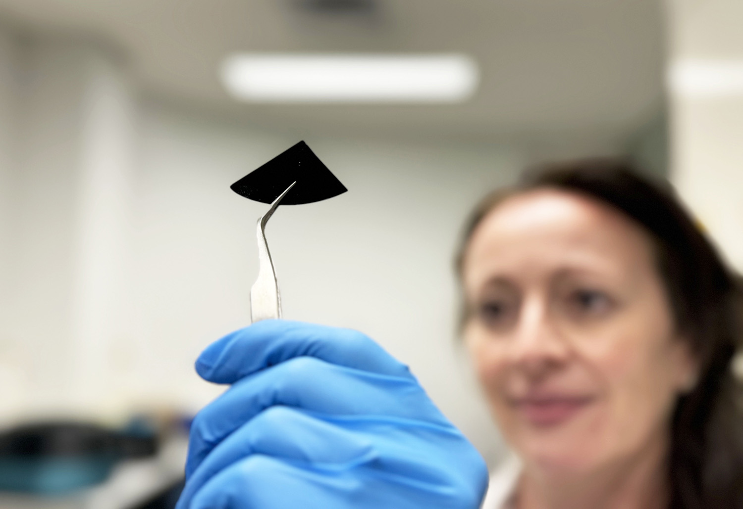
Spike the viruses to kill them
Corresponding author Dr Natalie Borg, from RMIT’s School of Health and Biomedical Sciences, said this seemingly unsophisticated concept of skewering the virus required considerable technical expertise.
“Our virus-killing surface looks like a flat black mirror to the naked eye but actually has tiny spikes designed specifically to kill viruses,” she said.
“This material can be incorporated into commonly touched devices and surfaces to prevent viral spread and reduce the use of disinfectants.”
The nano spiked surfaces were manufactured at the Melbourne Centre for Nanofabrication, starting with a smooth silicon wafer, which is bombarded with ions to strategically remove material.
The result is a surface full of needles that are 2 nanometers thick – 30,000 times thinner than a human hair – and 290 nanometers high.
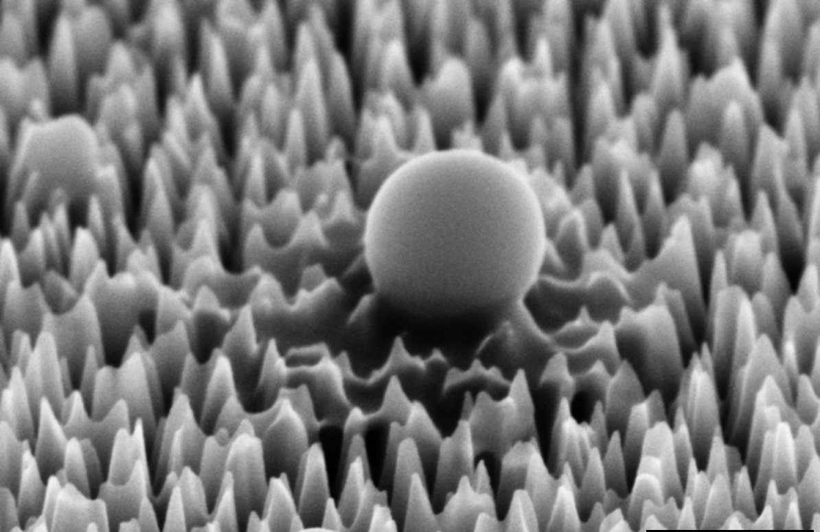
A virus on the nano spiked silicon surface, magnified 65,000 times. After one hour it has already begun to leak material.
Specialists in antimicrobial surfaces
The team led by RMIT Distinguished Professor Elena Ivanova has years of experience studying mechanical methods for controlling pathogenic microorganisms inspired by the world of nature: the wings of insects such as dragonflies or cicadas have a nanoscale spiked structure that can pierce bacteria and fungi.
In this case, however, viruses are an order of magnitude smaller than bacteria so the needles must be correspondingly smaller if they are to have any effect on them.
The process by which viruses lose their infectious ability when they contact the nanostructured surface was analysed in theoretical and practical terms by the research team.
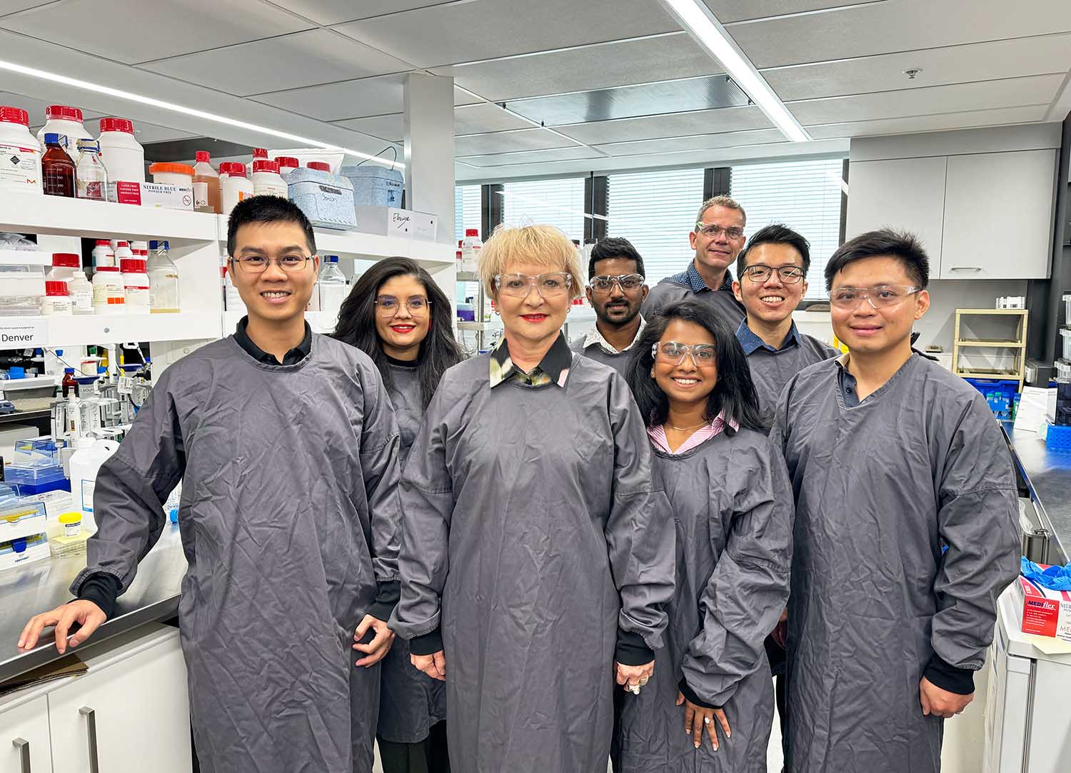
Team Ivanova with study corresponding author Professor Elena Ivanova (3rd from left) and study first author Samson Mah (2nd from right). Credit: RMIT.
Researchers at Spain’s Universitat Rovira i Virgili (URV), Dr Vladimir Baulin and Dr Vassil Tzanov, computer simulated the interactions between the viruses and the spikes.
RMIT researchers carried out a practical experimental analysis, exposing the virus to the nanostructured surface and observing the results at RMIT’s Microscopy and Microanalysis Facility.
The findings show the spike design to be extremely effective at damaging the virus’ external structure and piercing its membranes, incapacitating 96% of viruses that came into contact with the surface within six hours.
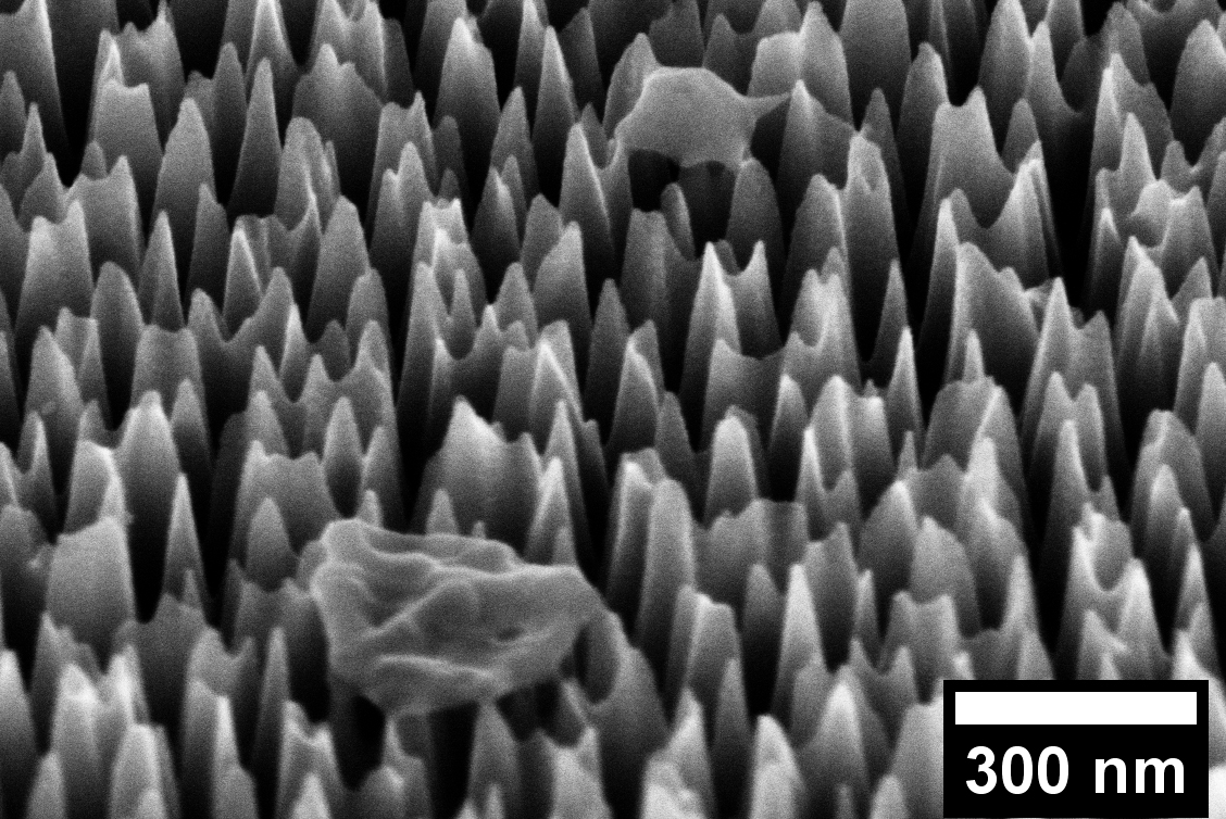
A virus on the nano spiked silicon surface, magnified 65,000 times. After six hours it has been completely destroyed. Credit: RMIT.
Study first author, Samson Mah, who completed the work under an RMIT-CSIRO Masters by Research Scholarship and has now progressed to working on his PhD research with the team, said he was inspired by the practical potential of the research.
“Implementing this cutting-edge technology in high-risk environments like laboratories or healthcare facilities, where exposure to hazardous biological materials is a concern, could significantly bolster containment measures against infectious diseases,” he said.
“By doing so, we aim to create safer environments for researchers, healthcare professionals, and patients alike.”
The project was a truly interdisciplinary and multi-institutional collaboration carried out over two years, involving researchers from RMIT, URV (Spain), CSIRO, Swinburne University, Monash University and the Kaiteki Institute (Japan).
The RMIT-CSIRO Masters by Research Program allows students to work with CSIRO and RMIT on a range of projects across science, engineering and health disciplines.
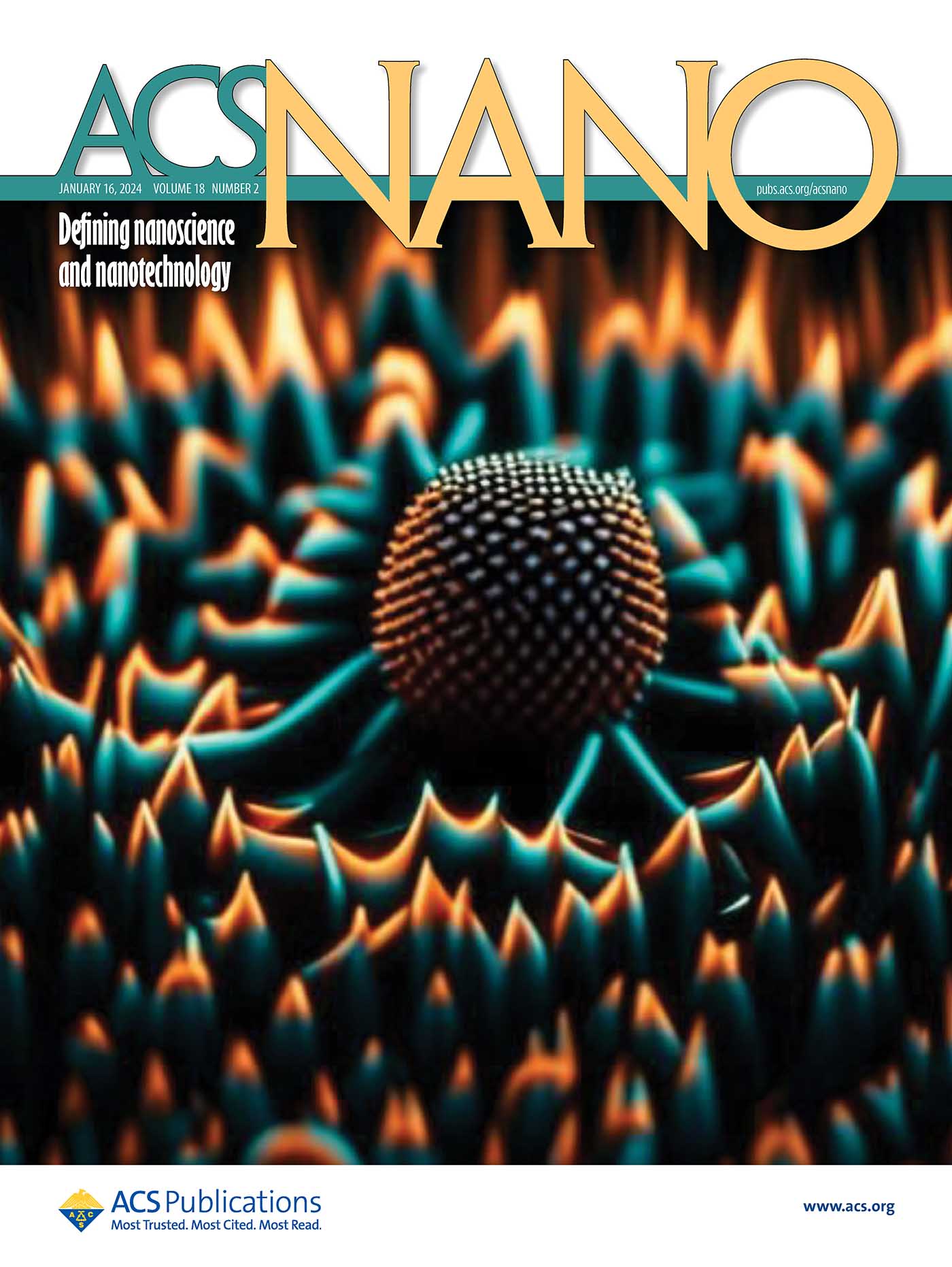
The team's AI generated image on the cover of the American Chemical Society's renowned nanotechnology journal ACS Nano.
This study was supported by the ARC Steel Research Hub and by the ARC Industrial Transformational Training Centre in Surface Engineering for Advanced Materials.
‘Piercing of the Human Parainfluenza Virus by Nanostructured Surfaces’ is published in ACS Nano. (DOI: 10.1021/acsnano.3c07099)
Story: Michael Quin with URV Science Communication and Outreach Unit.

